
Article
Open exposure for palatally impacted canines is an effective and well documented technique, with the tooth erupting into the palate relatively quickly, and usually without complication.1 On occasion, it may be necessary to bond an attachment soon after surgery, either to prevent the mucosa re-covering the tooth or to commence alignment. Typically, the ideal position for bonding the attachment is close to the gingival margin due to partial eruption and rotation of the canine.
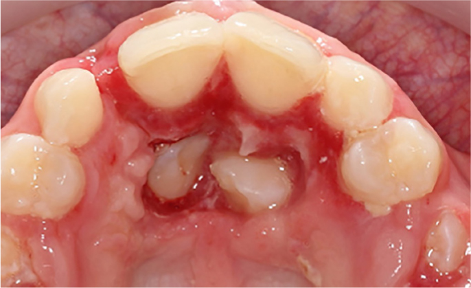
Difficulty in maintaining good oral hygiene in the immediate post-surgical period means that the mucosa and gingivae are often inflamed and prone to bleeding (Figure 1).
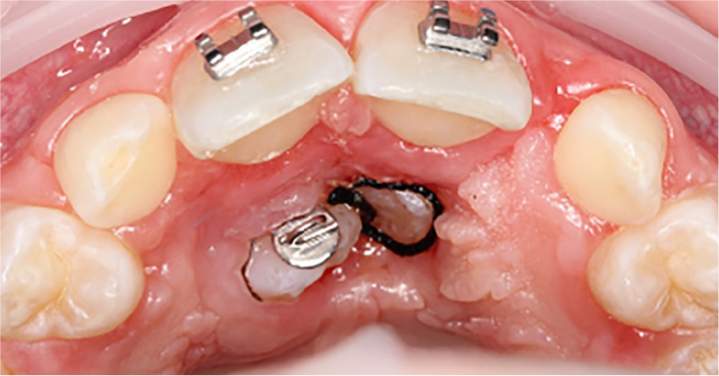
A method which greatly increases bonding success in situations where blood and gingival fluid are present is the placement of retraction cord soaked in styptic around the margin of the tooth. This provides simultaneous haemostasis and retraction of the soft tissues, increasing the surface area available for bonding and reducing contamination.
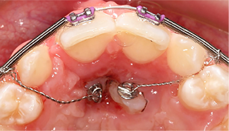
The tooth to be bonded should be cleaned and dried as much as possible and wide-diameter retraction cord, soaked in 15.5% ferric sulfate solution, gently packed into the sulcus (Figure 2). The retraction cord should be removed after 60 seconds and the tooth irrigated with water and air dried before application of the usual etchant, bonding agents and placement of an attachment (Figure 3).
This method is demonstrated in a 13-year-old female who underwent open exposure of both maxillary canines and inflammation of the surrounding mucosa. If cord is not available, retraction paste dispensed from a campule may be used.