Johnson K A study of the dimensional changes occurring in the maxilla following tooth extraction. Aust Dent J. 1969; 14:241-244
Atieh MA, Alsabeeha NH, Payne AG Interventions for replacing missing teeth: alveolar ridge preservation techniques for dental implant site development. Cochrane Database Syst Rev. 2015;
Hammerle CH, Araujo MG, Simion M Evidence-based knowledge on the biology and treatment of extraction sockets. Clin Oral Implants Res. 2012; 23:80-82
Thomas S, Turner SR, Sandy JR Autotransplantation of teeth: is there a role?. Br J Orthod. 1998; 25:275-282
Malmgren B Decoronation: how, why, and when?. J Calif Dent Assoc. 2000; 28:846-854
Sapir S, Shapira J Decoronation for the management of an ankylosed young permanent tooth. Dent Traumatol. 2008; 24:131-135
Mohadeb JV, Somar M, He H Effectiveness of decoronation technique in the treatment of ankylosis: a systematic review. Dent Traumatol. 2016; 32:255-263
Chae JM, Paeng JY Orthodontic treatment of an ankylosed maxillary central incisor through single-tooth osteotomy by using interdental space regained from microimplant anchorage. Am J Orthod Dentofacial Orthop. 2012; 141:e39-e51
Kofod T, Wurtz V, Melsen B Treatment of an ankylosed central incisor by single tooth dento-osseous osteotomy and a simple distraction device. Am J Orthod Dentofacial Orthop. 2005; 127:72-80
Uzuner FD, Darendeliler N Dentoalveolar surgery techniques combined with orthodontic treatment: a literature review. Eur J Dent. 2013; 7:257-265
Morgan C, Howe L The restorative management of hypodontia with implants: I. Overview of alternative treatment options. Dent Update. 2003; 30:562-568
Uribe F, Chau V, Padala S Alveolar ridge width and height changes after orthodontic space opening in patients congenitally missing maxillary lateral incisors. Eur J Orthod. 2013; 35:87-92
Uribe F, Padala S, Allareddy V, Nanda R Cone-beam computed tomography evaluation of alveolar ridge width and height changes after orthodontic space opening in patients with congenitally missing maxillary lateral incisors. Am J Orthod Dentofacial Orthop. 2013; 144:848-859
Beyer A, Tausche E, Boening K, Harzer W Orthodontic space opening in patients with congenitally missing lateral incisors. Angle Orthod. 2007; 77:404-409
McDowall RJ, Yar R, Waring DT 2 ‘2’ 1: Orthodontic repositioning of lateral incisors into central incisors. Br Dent J. 2012; 212:417-423
Hemmings K, Harrington Z Replacement of missing teeth with fixed prostheses. Dent Update. 2004; 31:137-141
King PA, Foster LV, Yates RJ, Newcombe RG, Garrett MJ Survival characteristics of 771 resin-retained bridges provided at a UK dental teaching hospital. Br Dent J. 2015; 218:423-428
Karoussis IK Long-term implant prognosis in patients with and without a history of chronic periodontitis: a 10-year prospective cohort study of the ITI Dental Implant System. Clin Oral Implants Res. 2003; 14:329-339
Papaspyridakos P, Chen CJ, Singh M, Weber HP, Gallucci GO Success criteria in implant dentistry: a systematic review. J Dent Res. 2012; 91:242-248
Derks J, Hakansson J, Wennstrom JL, Klinge B, Berglundh T Patient-reported outcomes of dental implant therapy in a large randomly selected sample. Clin Oral Implants Res. 2015; 26:586-591
Williams P, Travess H, Sandy J The use of osseointegrated implants in orthodontic patients: I. Implants and their use in children. Dent Update. 2004; 31:287-290
Thilander B, Odman J, Lekholm U Orthodontic aspects of the use of oral implants in adolescents: a 10-year follow-up study. Eur J Orthod. 2001; 23:715-731
Jivraj S, Chee W Treatment planning of implants in posterior quadrants. Br Dent J. 2006; 201:13-23
Jivraj S, Chee W Treatment planning of implants in the aesthetic zone. Br Dent J. 2006; 201:77-89
Tarnow DP, Magner AW, Fletcher P The effect of the distance from the contact point to the crest of bone on the presence or absence of the interproximal dental papilla. J Periodontol. 1992; 63:995-996
Tooth autotransplantation is a versatile and successful technique if used in suitable cases; however, it is not always the optimal treatment choice. This article will explore alternative treatment strategies for managing failing or missing teeth, including methods for managing the bone, orthodontic options and techniques for tooth replacement. These methods may be considered as an adjunct to tooth transplantation, or an alternative, if transplantation is not deemed appropriate. Indications for alternative treatments are discussed with illustrations from treated cases.
CPD/Clinical Relevance: A number of approaches are available for managing failing or missing teeth and are dependent on the clinical situation. It is important for dental specialists to understand these options and to work collaboratively to determine the best option for patients on an individual basis.
Article
Sophy Barber
The advantages of tooth autotransplantation and the broad applications for which the technique can be used have been outlined in the previous reports in this series. While tooth autotransplantation is a highly versatile and successful technique, it is not suitable for all cases with failing or missing teeth, and other management strategies may be preferable (Table 1). These alternative treatment options are described with an explanation of the purpose of the treatment and indications for use, with illustrations from clinical cases.
Purpose of treatment
Treatment modality
Indication for use
Preservation of alveolar bone
Decoronation
Management of an ankylosed tooth in a growing patient to prevent bony deficits developing.Used to preserve alveolar bone during dental development prior to definitive tooth replacement with tooth transplantation or prosthetic tooth.
Dento-alveolar osteotomy
Repositioning of an ankylosed tooth in a non-growing patient, usually undertaken as part of a comprehensive orthodontic treatment. Ankylosed tooth may eventually be lost and require replacement.
Alveolar ridge preservation techniques
Techniques used to preserve bone, usually for future placement of a dental implant. Methods include hard and soft tissue grafting with concomitant use of barrier membranes, usually as an adjunct to atraumatic tooth extraction.
Orthodontic tooth movement in edentulous site
Movement of tooth through edentulous site to encourage bone deposition to aid tooth replacement with tooth transplantation or prosthetic tooth.
Space elimination
Orthodontic space closure with camouflage
Movement of adjacent teeth into edentulous site to eliminate the need for tooth replacement. The substitute tooth and adjacent teeth can be camouflaged using restorative techniques to improve aesthetics.
Orthodontic space redistribution
Movement of adjacent teeth to change site requiring tooth replacement.
Tooth replacement
Removable prostheses
Interim treatment for temporary tooth replacement.Definitive treatment in cases where other treatment methods contra-indicated.
Tooth-supported prostheses
Temporary or permanent method of tooth replacement in growing or non-growing patients.
Implant-supported prostheses
Permanent method of tooth replacement in non-growing patients in sites with adequate space and bony volume.
Preservation of alveolar bone
Reduction in alveolar bone volume has been recognized as an undesirable sequelae to tooth loss for more than four decades.1 A lack of alveolar bone is problematic as it limits the options available for tooth replacement,2 and the associated gingival recession can cause additional problems for prosthodontic rehabilitation in the aesthetic zone.3 One of the greatest advantages of tooth autotransplantation is the ability of the donor tooth to preserve the height and volume of the alveolar bone in the recipient site.4 However, in cases where tooth transplantation is not a suitable treatment option, other methods of bone management may be considered. These methods include decoronation, dento-osseous osteotomy and alveolar ridge preservation techniques.
Decoronation
Decoronation is a popular method for preventing bone defects associated with infraocclusion that occurs secondary to ankylosis in growing patients. It is most commonly used for growing children who have undergone severe dental trauma in the anterior maxilla region. Decoronation involves a coronectomy to remove the crown of the ankylosed tooth. This needs to be extended below the level of the cemento-enamel junction and 1 mm under the crestal bone margin. Previous endodontic materials are removed through instrumentation of the pulp canal and saline is used for thorough irrigation. The aim is to induce bleeding and clot formation in the canal, providing cells for replacement resorption.5 The root of the ankylosed tooth is left in the bone with primary closure of the mucosa to encourage soft tissue healing and bone apposition.6 Replacement resorption of the root is expected to continue but with simultaneous bone deposition, resulting in minimal loss of alveolar bone. Reorganization of the transeptal fibres encourages bone growth in line with vertical alveolar development of adjacent teeth. Placing an interim removable or fixed tooth replacement into the coronal space following mucosal healing helps to maintain the space in the arch and restore aesthetics. Observational studies demonstrate that decoronation successfully maintains the alveolar bone and can even lead to a small gain in ridge height, although success depends on timing the procedure correctly.7 Decoronation is used as an interim treatment to maintain bone in growing patients whilst awaiting the necessary dental development for definitive treatment. Figure 1 illustrates the decoronation procedure, which was undertaken to manage progressive ankylosis of an incisor following trauma in a young patient. The bone was maintained in the site until the patient was ready for tooth autotransplantation.
Figure 1. Decoronation procedure for an ankylosed maxillary right central incisor with infraocclusion in a growing patient. (a) A buccal flap is raised to enable visualization of the tooth and supporting bone. (b) Coronectomy extending 1 mm under the crestal bone. (c) Pulp extirpation from the root canal in the remaining root and induction of intracanal bleeding. (d) Primary closure of the mucosal flap. (e) Post-operative healing. (f) The remaining root left in situ to maintain bone and maximize future options for tooth replacement.
Dento-osseous osteotomy
An alternative method for managing ankylosed, infraoccluded teeth involves repositioning a tooth or blocks of teeth within the surrounding bone using a dento-osseous osteotomy. This technique is most commonly used for a single tooth and subsequently is referred to as a single tooth osteotomy. The tooth is separated within a segment of bone, usually using a piezoelectric instrument to minimize damage to adjacent tooth roots. The fragment can then be repositioned in one of two ways. In cases where the movement is small, the segment can be placed immediately into the correct position and secured using fixed ligation.8 Alternatively, in cases where this is not feasible due to the extent of movement required, an orthodontic appliance can be used to move the segment into the correct position gradually using a distraction osteogenesis type technique with the ankylosed tooth as the point of force application.9 Bone healing is completed within four to six weeks and orthodontic post-surgical mobilization of the segment should therefore be completed within two to four weeks.10Figure 2 illustrates the use of single-tooth osteotomy to reposition an ankylosed incisor in a non-growing patient following dental trauma.
Figure 2. A single tooth dento-osseous osteotomy of a maxillary right lateral incisor. (a) Infraocclusion of the tooth secondary to ankylosis following trauma. (b) Periapical radiograph illustrating the ankylosed position of the maxillary lateral incisor and associated vertical defect in the alveolar bone. The canal obliteration of this tooth and endodontic treatment of the adjacent canine are further evidence of the trauma history. (c) The lateral incisor position at the time of surgery following fixed appliance treatment to align the maxillary arch. (d) Separation of the bone segment containing the lateral incisor. (e) Repositioning of the segment using an orthodontic bracket on the tooth and the archwire. (f) The tooth is secured to the archwire and the mucosa is sutured closed around the tooth.
Dento-osseous osteotomies are not without risks and the main concerns are loss of tooth vitality, avascular necrosis in the bone segment, gingival recession, loss of crestal bone and pocket formation, and a delay in movement of the segment due to bone interferences.10 Single tooth osteotomies are only suitable for patients where minimal further growth is expected, as the underlying ankylosis is not addressed, so the tooth will remain static following the cessation of orthodontic treatment. In addition, the process of replacement resorption will progress more rapidly in growing patients, leading to earlier loss of the ankylosed tooth and therefore less treatment benefit. Single tooth osteotomy technique provides a method for moving an ankylosed tooth within an orthodontic treatment plan. It allows other aspects of malocclusion to be resolved but it is not a definitive treatment. The eventual loss of the ankylosed tooth should be considered and the long-term plan is likely to involve tooth replacement.
Alveolar ridge preservation
Another suggested approach to bone management is alveolar ridge preservation, also called socket preservation or alveolar ridge grafting. This is an umbrella term for techniques that aim to maintain favourable alveolar ridge architecture for future tooth replacement. Alveolar ridge preservation techniques involve placement of a graft material alone, or in combination with a barrier membrane, at the time of tooth extraction, theoretically to encourage osteoconduction and osteoinduction. Alveolar ridge preservation has been widely reported in conjunction with implant-supported tooth replacement, but it is less likely to have a role in tooth transplantation as the donor tooth is usually transplanted at the same time as the failing tooth is extracted, eliminating the risk of alveolar resorption. A Cochrane review of eight RCTs evaluating alveolar ridge preservation techniques in adults found limited evidence to demonstrate their effectiveness in minimizing ridge changes,2 and no evidence could be found for growing patients.
Orthodontic tooth movement to develop bone
The alveolus is functional bone and therefore bone only develops and persists in the presence of teeth. Underdevelopment causing atrophic alveolar ridges or local bony deficits is most often reported in people with hypodontia11 and management through orthodontic movement of adjacent teeth through agenesis sites has been advocated as a method to increase bone volume prior to tooth replacement.12 This technique can theoretically be applied to any site with inadequate bone. However, the results from this are variable and bone resorption can occur if bone-maintaining methods of tooth replacement are not considered prior to orthodontic movement.13,14,15
Orthodontic management
Orthodontic space closure
All methods of tooth replacement, including tooth transplantation, carry a risk of failure that obligates patients to future dental treatment. For some people with existing or potential tooth loss, this commitment to long-term dental treatment may be insurmountable and, instead, they may wish to consider treatment planning options that obviate the need for tooth replacement. Most commonly this involves orthodontic space closure, where fixed appliances are used to move the adjacent teeth either to eliminate the need for tooth replacement completely or to relocate the edentulous space into a site that is more amenable to tooth replacement. The suitability of orthodontic space closure depends on the site of the missing tooth, the morphology of adjacent teeth and other aspects of malocclusion (Table 2). Elimination of maxillary lateral incisor, mandibular incisor and second premolar agenesis sites are the most common application for orthodontic space closure. Camouflage of maxillary lateral incisors in the central incisor position is challenging due to the discrepancy in crown and root widths and the subsequent difficulties in achieving a satisfactory functional and aesthetic restoration. Modifications in tooth positioning aid restoration and reduce the occlusal forces on the substitute tooth.16Figure 3 shows a patient who lost both central incisors through dental trauma. The treatment plan involved orthodontic space closure with the lateral incisors camouflaged as central incisors and the canines replacing the lateral incisors.
Considerations for all treatment methods
Patient wishes
Presenting complaintDesired outcomeCommitment to long-term maintenanceAbility to accept treatment
Dental health
Current health of teethPeriodontal conditionOral hygieneHistory of dental disease
Relevant medical history
Any medical factor that may impact on ability to accept treatment
Patient's age and maturity
Growing or non-growingUnderstanding of treatment options
Occlusion
Crowding or spacingOther missing teethInter-arch relationship
Considerations specifically for tooth replacement
Size of edentulous space
Suitability for restorationScope for optimizing space
Health and position of adjacent teeth
Suitability to act as abutments
Recipient site factors
Space in archAlveolar bone volume and qualityPosition of adjacent tooth roots
Gingival architecture
Impact on potential restoration
Functional occlusion
Potential interferences Occlusal loading
Considerations specifically for space closure
Amount of space closure required
Impact of closing space on occlusionFeasibility of space closureNeed for anchorage support
Morphology of substitute tooth
Crown size and shapeEmergence profile
Morphology of adjacent teeth
Ability to achieve harmony and symmetry with or without camouflage techniques
Gingival architecture
Impact on potential restoration
Figure 3. Orthodontic space closure with substitution of the maxillary lateral incisors into the position of the avulsed central incisors. (a) The patient presented for orthodontic assessment prior to suffering the dental trauma, in which both central incisors were avulsed. Initially the treatment plan involved extraction of maxillary premolars to create space for alignment. (b) Following avulsion of the central incisors, the plan was modified. It was agreed that the lateral incisors would be used to replace the central incisors, accepting the canines as substitxsutes for the lateral incisors and premolars in the canine position. (c) Removal of the fixed appliance and chairside restoration of the lateral incisors by the treating orthodontist. This was an interim measure to allow the gingival inflammation to resolve prior to gingival recontouring and definitive restorative treatment to camouflage the anterior four teeth.
Orthodontic space redistribution
Space redistribution can be used to move sound teeth with good root length and morphology into sites which provide a good occlusal, restorative, aesthetic and functional result. For example, in a case where canines and second premolars are missing, the first premolar can potentially be mesialized into the canine position. Restoration of the canine region is highly unpredictable with limited availability of successful options, compared to the premolar sites, which have lower aesthetic and occlusal demands and more treatment options from adjacent strategic teeth.
Methods of tooth replacement
Tooth transplantation is one method of tooth replacement, but in cases where a donor tooth is not available, other methods of tooth replacement may be considered. For replacement of one or two teeth, these treatment options fall into three categories:
Removable mucosal or tooth-borne prostheses;
Tooth-supported fixed prostheses; and
Implant-supported fixed prostheses.
1. Removable prostheses
Removable prostheses are usually considered in the interim, rather than as a definitive method of tooth replacement in young people where only one or two teeth are missing. The prostheses rely on the soft tissues or the dentition for support and retention. However, in certain cases, removable prostheses may be the definitive treatment option if other methods of tooth replacement are contra-indicated; for example, in those with complicated risk factors or medical health issues, long edentulous spans and poorly strategic adjacent teeth. The prosthesis of choice is most commonly an acrylic denture, although cobalt chrome dentures and orthodontic retainer type designs may also be considered. Removable prostheses have a number of advantages: replacement of hard and soft tissue defects,11 appliance removal to facilitate effective oral hygiene, relatively cheap and easy to manufacture, capacity for adjustments and incorporation of additional features. Removable prostheses can take some time to adjust to and compliance in young children is variable.
2. Tooth-supported fixed prostheses
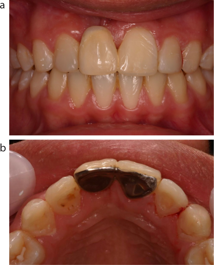
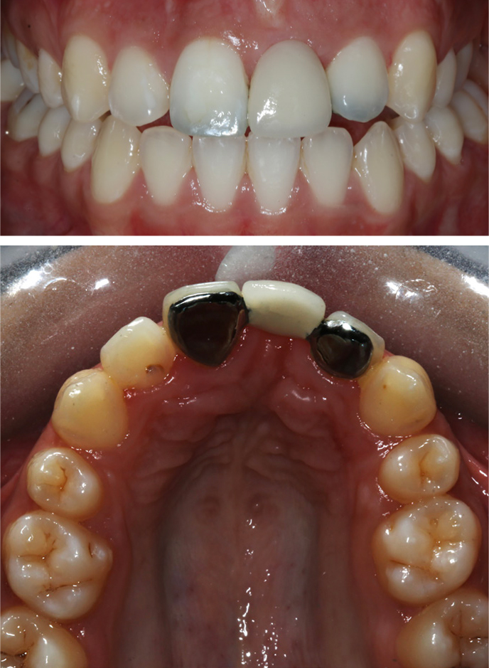
The two main types of tooth-supported fixed prostheses commonly considered for replacing one or two missing tooth units are resin-bonded bridges and conventional bridges.17 Resin-bonded bridges (RBB) with either a single wing cantilever design or a double wing fixed-fixed design are the most popular type of tooth-supported fixed prostheses. Single wing cantilever designs are preferred by many restorative dentists as there is concern that bond failure of one wing in a fixed-fixed design may result in undetected caries under the debonded wing. In the RBB design, a ceramic pontic is attached to adjacent teeth via a non-precious wing or wings, using enamel bonding adhesives. The main advantage of RBBs is that no or minimal preparation is required to facilitate placement. Estimated 10-year survival rates are 80.4% (95% confidence interval 77.6–83.2%).18 Clinical variables influencing survival revealed that design of the restoration, consideration of the occlusion, cementation technique and experience of the operator were significant factors. Minimal tooth preparation was shown to be superior for longevity compared with other types of preparation. Patient satisfaction has been shown to be high for this type of treatment. This makes treatment simple and predictable to deliver, with or without anaesthetic, and with no biological cost to the patient. For young patients, RBBs can be attached with provisional cement, allowing removal at a later date, if necessary, making them a suitable temporary measure for tooth replacement. The pontic is able to deliver excellent aesthetics, and improvements in opaque luting cements have reduced the problems caused by grey discoloration as a result of shine-through from the metal wing.17Figures 4 and 5 show patients who underwent tooth replacement with a RBB, as autotransplantation was contra-indicated due to the lack of a suitable donor tooth.
Figure 4.
(a, b) Placement of a resin-bonded bridge for a patient with a poor prognosis right maxillary central incisor following dental trauma. The crown of the failing tooth was used to create the pontic by connecting the extracted crown to the adjacent tooth via butterfly metal wings. This provides an ideal match in terms of morphology and shade.Figure 5.
(a, b) Placement of a resin-bonded bridge by the patient's general dentist to replace the maxillary left central incisor in a patient who did not want to undergo tooth autotransplantation.
Conventional bridges are less commonly used due to the need for extensive tooth preparation of the abutment teeth to provide sufficient coronal reduction for placement of a full-coverage abutment. Conventional bridges are indicated for patients who have existing coronal coverage restorations in suitable abutment teeth, where further preparation would not be deemed to be destructive and a full coverage restoration would be beneficial.
3. Implant-supported tooth replacement
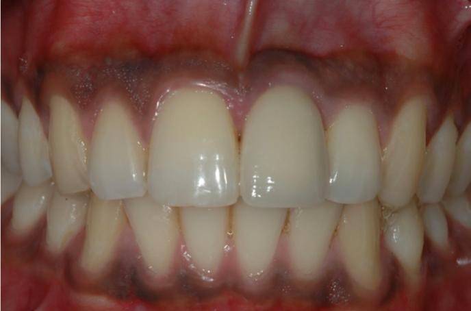
Implant-supported tooth replacement has gained widespread popularity over the last 40 years. The survival rate of implants in adults has been shown to be as high as 96.5% over 10 years,19 although success is more difficult to estimate due to wide variation in the criteria used to evaluate success20 and heterogeneity in treatment protocols. Patient-reported outcomes from implants, such as satisfaction with appearance and function, have been demonstrated to be high in well planned cases with good bone quantity and favourable clinical factors.21 Implant success is dependent on osseointegration to gain stability and this fusion to the bone prevents the implant-supported structure adapting and erupting with the surrounding teeth. As such, implants are not advocated for use in growing patients.22 Evidence of infraocclusion of up to 1.6 mm over 3 years has been demonstrated in anterior implants placed in adolescents.23 In those where growth is complete, additional factors associated with the recipient site need to be assessed to determine the feasibility of implant treatment. Implants require adequate bone volume for placement to allow for a functional and aesthetic result. Ideally, a distance of 1.5–2.0 mm of interseptal bone is required between the implant and adjacent roots to encourage papilla regeneration.24 Augmentation procedures may be possible to manage horizontal and some vertical and bucco-lingual deficits; however, inadequate mesio-distal space between the adjacent tooth crown and roots may prevent their use. Sufficient coronal space is required to allow a restoration that mimics the natural contours and is in harmony with the surrounding teeth.25 The distance between the contact point and alveolar crest has been shown to be highly important to papilla presence.26 Lack of mesio-distal space and bone quality are the most common contra-indications to implant use in maxillary lateral incisor and mandibular incisor sites. Figure 6 shows implant-supported tooth replacement in an adult who did not wish to undergo tooth autotransplantation.
Figure 6. A single screw-retained implant crown replacing the left maxillary central incisor in an adult patient. This tooth was lost following trauma and the resulting buccal bone concavity required simultaneous bone augmentation at the time of implant placement.
Conclusions
Tooth transplantation is a highly successful and biological method of tooth replacement. The outcome may be improved by the use of adjunctive treatments, such as decoronation and temporary tooth replacement, to allow transplantation to be delayed until the optimum time. This paper highlights the importance of understanding alternative treatment options of tooth replacement and the advantages and disadvantages of each.