Hanisch M, Hanisch L, Kleinheinz J, Jung S. Primary failure of eruption (PFE): a systematic review. Head Face Med. 2018; 14
Rhoads SG, Hendricks HM, Frazier-Bowers SA. Establishing the diagnostic criteria for eruption disorders based on genetic and clinical data. Am J Orthod Dentofacial Orthop. 2013; 144:194-202
Sharma G, Kneafsey L, Ashley P, Noar J. Failure of eruption of permanent molars: a diagnostic dilemma. Int J Paediatr Dent. 2016; 26:91-99
Proffit WR, Vig KWL. Primary failure of eruption: a possible cause of posterior open-bite. Am J Orthod. 1981; 80:173-190
Baccetti T. Tooth anomalies associated with failure of eruption of first and second permanent molars. Am J Orthod Dentofacial Orthop. 2000; 118:608-610
Ahmad S, Bister D, Cobourne MT. The clinical features and aetiological basis of primary eruption failure. Eur J Orthod. 2006; 28:535-540
Raghoebar GM, Boering G, Vissink A, Stegenga B. Eruption disturbances of permanent molars: a review. J Oral Pathol Med. 1991; 20:159-166
Frazier-Bowers SA, Koehler KE, Ackerman JL, Proffit WR. Primary failure of eruption: further characterization of a rare eruption disorder. Am J Orthod Dentofacial Orthop. 2007; 131:578.e1-11
Frazier-Bowers SA, Simmons D, Wright JT, Proffit WR, Ackerman JL. Primary failure of eruption and PTH1R: the importance of a genetic diagnosis for orthodontic treatment planning. Am J Orthod Dentofacial Orthop. 2010; 137:160.e1-7
Decker E, Stellzig-Eisenhauer A, Fiebig BS, Rau C, Kress W, Saar K, Ruschendorf F, Hubner N, Grimm T, Weber BH. PTHR1 loss-of-function mutations in familial, nonsyndromic primary failure of tooth eruption. Am J Hum Genet. 2008; 83:781-786
Mubeen S, Seehra J. Failure of eruption of first permanent molar teeth: a diagnostic challenge. J Orthod. 2018; 45:129-134
Suri L, Gagari E, Vastardis H. Delayed tooth eruption: pathogenesis, diagnosis, and treatment. A literature review. Am J Orthod Dentofacial Orthop. 2004; 126:432-445
Kjær I. Mechanism of human tooth eruption: review article including a new theory for future studies on the eruption process. Scientifica. 2014;
Yildirim D, Yilmaz HH, Aydin U. Multiple impacted permanent and deciduous teeth. Dentomaxillofac Radiol. 2004; 33:133-135
Nagpal A, Sharma G, Sarkar A, Pai KM. Eruption disturbances: an aetiological-cum-management perspective. Dentomaxillofac Radiol. 2005; 34:59-63
Sivakumar A, Valiathan A, Gandhi S, Mohandas AA. Idiopathic failure of eruption of multiple permanent teeth: report of 2 adults with a highlight on molecular biology. Am J Orthod Dentofacial Orthop. 2007; 132:687-692
Blechman AM, Smiley H. Magnetic force in orthodontics. Am J Orthod. 1978; 74:435-443
Sandler JP. An attractive solution to unerupted teeth. Am J Orthod Dentofacial Orthop. 1991; 100:489-493
This paper describes primary failure of eruption and presents some of the theories about the aetiology of this clinical condition. It also covers single ankylosed teeth as well as cases that present with multiple unerupted teeth. The various approaches to the clinical management of this not uncommon problem are discussed, along with the pros and cons of some of these techniques.
One difficult clinical challenge is documented, where a 14-year-old patient presented with 19 unerupted permanent teeth and, with the help of rare-earth magnets and upper and lower fixed appliances, within a two-year period a good result was achieved.
CPD/Clinical Relevance: Clinicians encounter teeth that have failed to erupt on a regular basis. Appropriate diagnosis and treatment planning of these cases, and subsequent effective clinical management, is imperative to ensure the most favourable outcome for our patients.
Article
Permanent teeth may fail to erupt because of obstruction, or disruption, of the eruptive mechanism. Eruption may be obstructed by the presence of pathology, ectopic tooth position, interferences from adjacent teeth or lateral forces from the tongue.1 Teeth may also fail to erupt due to primary failure of eruption (PFE) or ankylosis. The latter is defined as the fusion of cementum to bone in at least one area lacking a periodontal ligament space.2
Primary failure of eruption (PFE) is defined as incomplete tooth eruption despite the presence of a clear eruptive pathway.1 There is no ankylosis and it is the eruptive mechanism itself that is disturbed.2 This article will review the literature on PFE and failure of eruption from ankylosis. We also present a case of multiple unerupted teeth, treated with the use of neodymium iron boron magnets, as well as the more conventional deployment of gold chains to facilitate orthodontic traction.
Establishing the correct diagnosis forms the basis of satisfactory management of unerupted teeth. It is vital to distinguish obstructive failure of eruption, PFE and isolated ankylosis. Not doing so may jeopardize successful orthodontic management and potentially cause harm to the patient. This is particularly pertinent in cases of PFE, where the injudicious application of traction may precipitate ankylosis of the offending tooth and consequent intrusion of adjacent normal teeth.3
Primary failure of eruption
The term PFE was coined by Proffit and Vig in their seminal research on the topic.4 It is a rare condition with a prevalence of 0.06%.5 Subsequent research has refuted some of their initial observations, but the literature demonstrates consensus on the following features:1,2,6
Posterior teeth are more frequently affected than anteriors;
Teeth posterior to the most anteriorly affected tooth may be involved;
Affected teeth may completely fail to erupt, or may initially erupt through the oral mucosa, before ceasing to erupt further;
The occlusion manifests as a lateral open bite;
Involvement can be unilateral or bilateral;
Application of orthodontic forces to the affected teeth sometimes precipitates ankylosis rather than normal tooth movement;
PFE is associated with a mutation in the parathyroid hormone 1 receptor (PTH1R) gene.
Raghoebar et al subdivided PFE into primary and secondary retention; primary if the tooth failed to erupt, and secondary if there was cessation after initial penetration through the oral mucosa.7 Frazier-Bowers et al described three different forms of PFE:8
Type I, where all affected teeth have a similar lack of eruptive potential and a posterior open bite establishes with worsening severity from anterior to posterior;
Type II, where a more varied eruption potential is seen between the affected teeth. In such cases, a tooth distal to the most mesial affected tooth may show greater, but still inadequate, eruptive potential;
In Type III, subjects have both Type I and Type II tendencies co-existing in different quadrants.
Several systemic or syndromic conditions, such as cherubism and cleidocranial dysplasia, have failure of tooth eruption as an identifying feature, and must be excluded when establishing a diagnosis of PFE.1 A family history of PFE appears to be a risk factor for its development, and inheritance appears to be autosomal dominant with variable expressivity.9 Earlier research found the level of dental anomalies, such as hypodontia, to be considerably higher than average in individuals with PFE, but more recent research refutes this.1,6
PFE appears to be associated with a mutation in the PTH1R gene and, consequently, genetic testing may assist in early and accurate diagnosis.10
Even in the absence of a known genetic, pathological or environmental factor responsible for failure of tooth eruption, a true definitive diagnosis of PFE may only ever be given retrospectively, after attempts at orthodontic extrusion of the affected teeth have failed. Management of PFE is made difficult by the tendency for affected teeth to ankylose when orthodontic forces are applied.1 The literature describes various techniques from coronal build-ups of the affected teeth to segmental osteotomy, but treatment in severe cases is invariably complex and often multidisciplinary.3 The case presented here responded well to orthodontic traction, excluding a diagnosis of PFE.
Ankylosis and failure of eruption
Isolated ankylosis is a rare condition with a similar presentation to PFE. A diagnostic feature distinguishing between the two is that ankylosis typically affects a single tooth, with distal teeth being unaffected.1 This naturally makes diagnosis difficult in a child in the mixed dentition, as one cannot be certain of the status of the unerupted teeth. Partially erupted teeth that are ankylosed will exhibit a dull metallic sound when percussed. These teeth cease to erupt, drift or move, despite normal adolescent growth or orthodontic traction. They may further disturb the occlusion by allowing adjacent teeth to tilt and opposing teeth to overerupt. Radiographically, ankylosis gives the appearance of relative submergence, and the periodontal ligament space may be focally absent.3,11,12
The orthodontic management of ankylosis differs significantly from that of primary failure of eruption. Isolated ankylosis responds well to treatment and may be managed by extracting the affected tooth at the appropriate age, or through luxation and subsequent orthodontic alignment.3,12
Multiple unerupted teeth
Delayed tooth eruption (DTE) is defined as the emergence of a tooth into the oral cavity at a time that deviates significantly from the norms established for different races, ethnicities and gender.11 The mechanisms and biological underpinnings of tooth eruption are presently poorly understood, however, several rare diseases and syndromes are associated with delayed tooth eruption.13
Occasionally, failure of eruption of multiple teeth cannot be attributed to a local or systemic condition. This is a rare occurrence and the literature contains relatively few examples of such cases. Their orthodontic management is varied, and differing approaches, applied with varying levels of success, have been reported.14,15,16
Orthodontic application of magnets
Many options are available for the management of unerupted teeth and these vary in their invasiveness and ease of application. The least invasive is the creation of space to facilitate spontaneous eruption, and the next line is surgical exposure of the unerupted tooth with attachment of a gold chain to allow application of orthodontic traction. This latter approach is probably the most common approach to impacted teeth, but it does require a surgical procedure, usually under general anaesthetic, and the operator must provide treatment that is relatively technique sensitive.
Blechman and Smiley first described the use of magnets to achieve orthodontic tooth movement, and since then magnets have been used successfully in a wide variety of orthodontic applications.17,18 An application for impacted teeth was first described by Sandler et al and involved the attachment of paralene coated, rare earth magnets to the unerupted tooth using composite resin, followed by provision of a removable appliance containing a larger magnet.19 The magnets must be correctly placed with opposing poles approximated, and properly aligned to ensure optimal direction of pull. Once they are in place, the only adjustment required is the occasional repositioning of the magnet contained within the removable appliance, until the two magnets are almost in apposition.
This method requires little manual dexterity of either the operator or the patient. The speed of tooth movement appears to be as quick as other methods, and the slowly increasing, continuous force may be more physiological. Palatally directed forces are more readily applied, helping to ensure that teeth do not erupt through the labial plate, and that they end up with an adequate cuff of attached gingiva on eruption.
Case report
A 14-year-old female patient presented with delayed eruption of multiple teeth. Medically she was fit and well and presented with a caries-free mixed dentition and excellent oral hygiene. Her initial complaint was that she was extremely anxious about the delayed eruption of her teeth, and had difficulty chewing on the left-hand side. There was no relevant family history and a genetic aetiology had not been identified.
Diagnosis
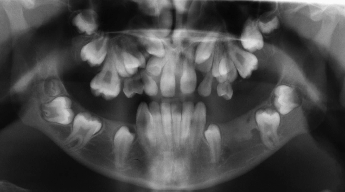
A panoramic radiograph (Figure 1) revealed the presence of 19 unerupted permanent teeth. The consultant in dental radiology reporting the OPT said that he could understand why some teeth had not erupted. He noted that the ‘bone on the left side of the mandible looked a bit dense’, however, did not suspect osteopetrosis. The patient also saw a consultant of paediatric bone care who noted that there were no concerns regarding her skeleton.
Figure 1. OPT showing multiple unerupted teeth.
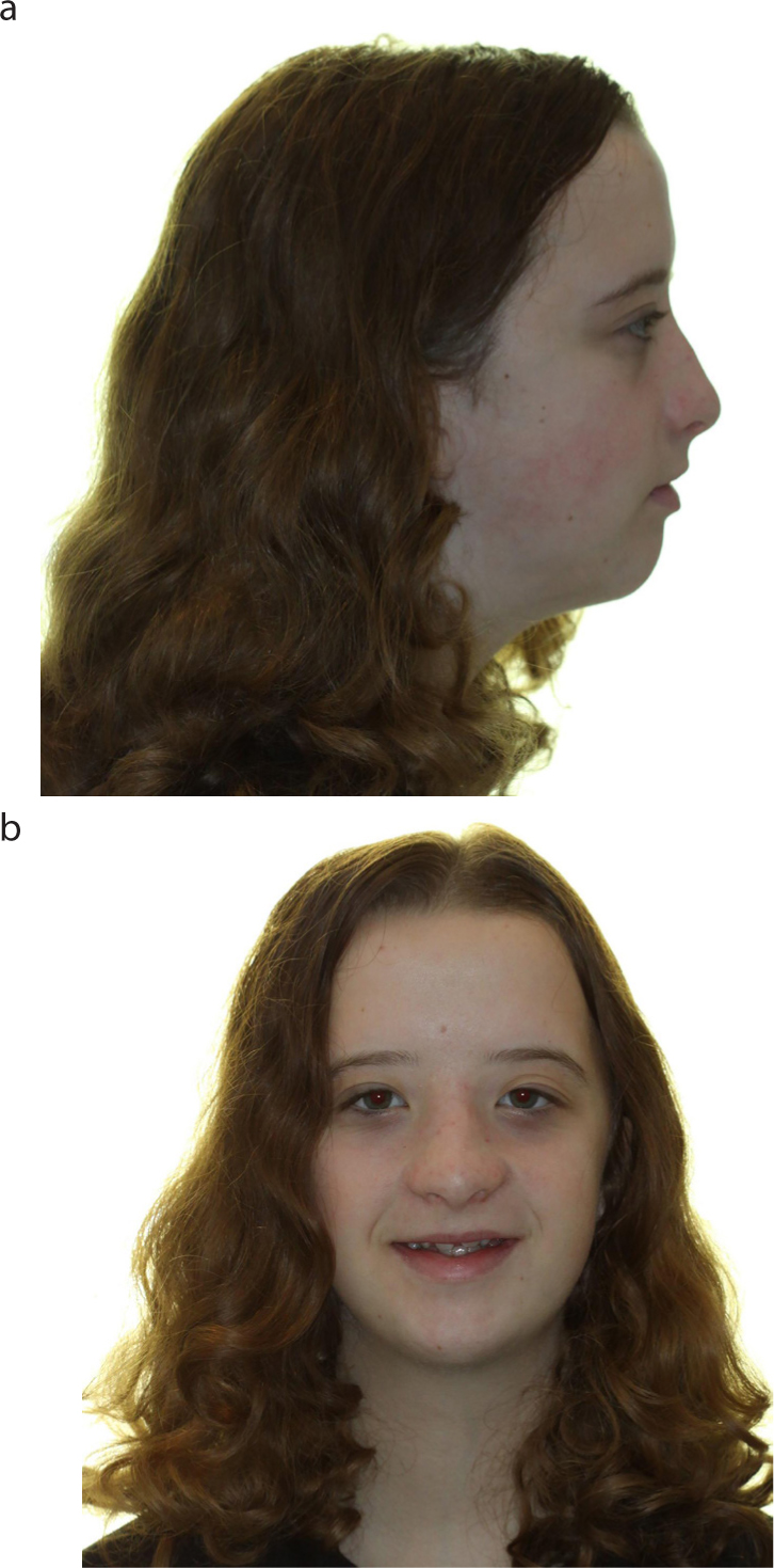
Dysmorphic features (Figure 2a, b), such as low set ears, had raised concerns in the past and she was under review by a geneticist. However, no specific diagnosis was made. It had been noted that she had hypertelorism, a small mandible, slightly high palate and hypermobile wrists. There was no evidence of cranial nerve compression and fundoscopy was unremarkable.
Figure 2.
(a, b) Dysmorphic features of hypertelorism and a small, retropositioned mandible.
After seeing a Consultant in Clinical Genetics, the patient enrolled into a research study called Deciphering Developmental Disorders (DDD). The initial report from that study said ‘No plausible pathogenic variants currently identified’.
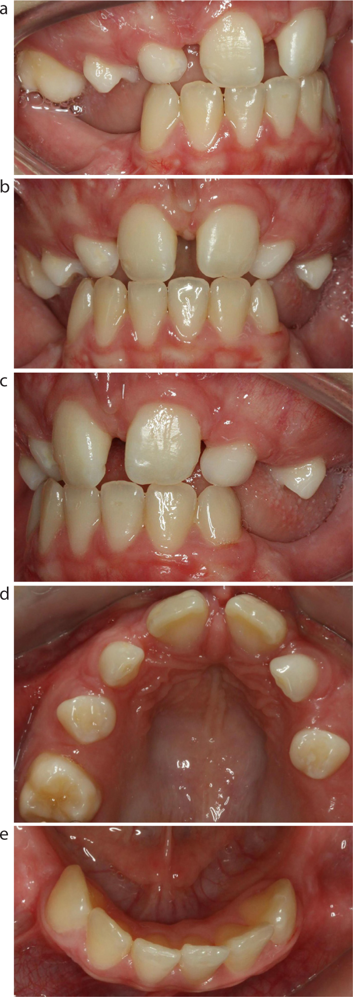
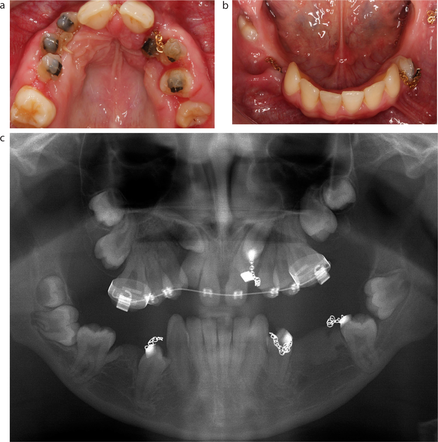
The orthodontic diagnosis was made of Class II incisor relationship on a mild Class II skeletal base with slight maxillary hypoplasia, and mandibular retrognathia, increased MMPA and lower facial height. She was still in the mixed dentition, with retained upper Cs and Ds and multiple unerupted teeth suffering from primary failure in eruption (Figures 3a–e).
Figure 3.
(a–e) Intra-oral views from initial presentation.
Treatment plan
The patient was referred to Chesterfield Royal Hospital for orthodontic treatment and a plan was devised to encourage her unerupted teeth into occlusion. This included extraction of remaining deciduous teeth, placement of an upper removable appliance containing rare earth magnets, and bonding of opposite pole magnets to the upper unerupted teeth to encourage eruption.
Treatment
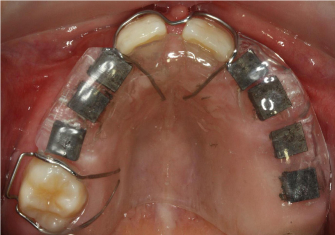
Orthodontic treatment was carried out over a 20-month period. Under general anaesthetic the remaining deciduous teeth, upper Ds and Cs, were removed and Neodymium Iron Boron magnets (3 x 3 x 1 mm) were bonded to UL542 and UR2456. Gold chains were bonded to the upper left canine, to the lower first premolars and to the lower left molar, to aid their eruption by eventually applying direct traction to the fixed appliances. An upper removable appliance was fabricated containing the larger magnets (5 x 5 x 2 mm) (Figure 4). Magnetic forces were applied for 7 months until the unerupted teeth were in a position where attachments could be placed to allow the direct orthodontic traction (Figure 5a–c).
Figure 4. Rare earth magnets embedded in an upper removable appliance.Figure 5.
(a, b) Unerupted teeth in a more favourable position for bonding. (c) OPT showing progress 9/12 into treatment.
The case was re-assessed at this stage with the aid of a CT scan which showed clear root damage to the UR2 from the unerupted UR3. It was therefore decided that the UR2 should be sacrificed along with the UL3. During the surgery, the surgeons also removed a triangle of bone mesial to the LL7 to aid eruption and exposed the UR7.
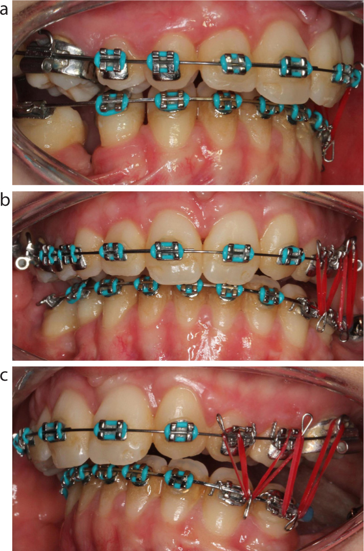
A routine course of orthodontics now followed involving upper and lower fixed appliances and attempted correction of the lateral open bite on the left with red elastics to Kobyashi hooks. Sadly, these teeth proved to be resistant to eruption (Figures 6a–c). An orthognathic opinion had been sought during treatment. It was decided that, although BSSO advancement would theoretically be possible, the risks of surgery were high due to thin mandible and uncertain quality of the bone. Lower second and third molars were also present, removal of which would be required prior to any osteotomy.
Figure 6.
(a–c) Upper and lower SWA elastics to attempt open bite closure.
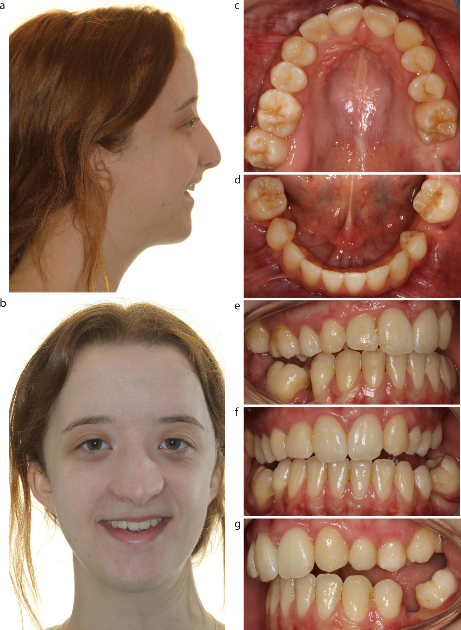
It was decided that the treatment had been largely successful, although it was impossible to achieve full closure of the lateral open bite. Despite this, the patient was completely happy with the results and declined any further intervention. She was, however, given the option of reconsidering further treatment in the future.
Three years after the end of treatment, the patient was still delighted with the result achieved, was really pleased with her smile and had no wish for further intervention (Figures 7a–g).
Figure 7.
(a–g) Long term result – 3 years after debond.
Conclusions
There is a variety of causes for unerupted teeth and it is helpful for all practitioners to be familiar with the various aetiological factors. It is certainly helpful if they can distinguish between primary failure of eruption and ankylosis.
Many different treatment approaches have been advocated for the management of unerupted teeth. In cases of multiple unerupted teeth, it is worthwhile considering using rare earth magnets, as multiple tooth movements can be carried out simultaneously to bring the teeth to a point where they can be bonded with conventional appliances and aligned.