Van der Linden FP, Bolender C, Canut JA. Three years postgraduate programme in Orthodontics. The final report of the Erasmus project. Eur J Orthod. 1992; 14:85-94
Ditmarov A. Orthodontics: Orthodontics vs Orthodontiya (Letter to the editor). Br Dent J. 2018; 2
Recognizing differences in the standards of orthodontic postgraduate education across Europe, the European Board of Orthodontists was established in 1997 with the aim of providing a standard against which an orthodontist could be judged independent of national barriers and, in so doing, promoting high standards of care. This article describes the experiences of both authors who sat the assessment at the June 2018 diet held at the European Orthodontic Congress in Edinburgh.
CPD/Clinical Relevance: Application for membership to the European Board of Orthodontists requires clinicians to work towards illustrating well-described and applied clinical procedures to correct significant orthodontic problems to an identified excellent standard. In the pursuit of attaining this recognition, orthodontists will invariably reflect on the treatments they provided and undoubtedly learn more, as well as seeing improvements in the standard of their patients' care.
Article
The American Board of Orthodontists (ABO) certification was created in 1929, with the aim of giving the orthodontist a clear view about the quality of care delivered and aiding the public in identifying quality-oriented orthodontists. In Europe, the development of the whole orthodontic specialty has been more haphazard, but with the adoption of a standard curriculum by many European universities, the European Orthodontic Society initiated the European Board of Orthodontists (EBO) in 1997.1 As recently as July 2018, there was a letter written to the British Dental Journal highlighting the problems in Russia with ‘Orthodontics vs orthodontiya’. The author suggested that referring dentists refer to board certified specialists as a means of ensuring that the orthodontic treatment provided would be performed in line with European approved protocols.2
In this article, two recently successful candidates at different stages of their careers will describe both routes to membership of the EBO.
Provisional EBO membership – Catherine
I am currently a second year post-CCST in Chesterfield and Sheffield. I started considering applying for provisional membership following some gentle encouragement from one of my supervising consultants after my MOrth (June 2016). The first examination I could have been eligible for was in June 2017, however, because of maternity leave I applied for the examination sitting in June 2018 in Edinburgh (the application deadline usually being the 31 December of the year prior to the EOS Congress at which you wish to be examined).
Over the last few years EOS membership has increased in numbers. There are, however, relatively fewer UK members compared to, for example, our Italian counterparts3,4 (Table 1).
Country
2001 Full
2018 Full (and Provisional Membership)
Austria
7
11 (8)
Belgium
3 (3)
Brazil
1
Bulgaria
(1)
Cyprus
1
Denmark
2
France
2
4 (4)
Germany
5
7 (2)
Greece
1
1 (1)
Iceland
1
Iran
2
Ireland
1
1
Italy
15
52 (8)
Japan
11
Lebanon
1 (1)
Monaco
1
1
Netherlands
2
2 (1)
Norway
(1)
Poland
2
Portugal
1
1 (1)
Saudi Arabia
1
Slovenia
(1)
Spain
5
15
Sweden
1
2
Switzerland
5
9 (8)
Turkey
1 (1)
UK
1
4(3)
United Arab Emirates
1
USA
1
TOTAL
48
137 (43)
Reasons for applying for provisional EBO membership
I think a few of my colleagues wondered ‘why on earth’ I decided to sign up for a seemingly unnecessary examination. My thoughts about applying for provisional membership were:
Some of my most interesting orthodontic conversations have been with European colleagues whom I have met at other meetings. I thought that membership to the European Society might facilitate better links with these colleagues to fuel more interesting debates and to share ideas about clinical techniques.
The examination would afford me an opportunity to measure myself against my European counterparts in a ‘spirit of self-improvement’.
I was extremely anxious about how I would manage my ISFE alongside my relatively new demands of being a mum. I was very uncertain about how I would manage the revision and stress. The EBO exam helped in two ways. The first was that I was able to cover all of the ‘basic MOrth’ orthodontic material 10 months before my ISFE, paving the way for me to revise more complex topics over the coming months. The second was that it provided some reassurance that my family and I could manage the ‘stress’ of an examination.
Preparation for examination
The guidelines outlining all eligibility criteria are clearly listed on the EOS website.5 The criteria have changed somewhat since the publication in 2003 by Sandler and Duterloo.4 Previously, candidates were usually established orthodontic specialists with at least 5 years of independent clinical experience. Since 2013, the eligibility criteria have changed to allow a period of provisional membership. Candidates are required to apply within 24 months of finishing their specialist training in orthodontics.
My preparation for the examination included:
Six weeks revision;
Approximately 30 hours for the two case write-ups and models to be prepared. The guidelines (https://www.eoseurope.org/ebo/EBOBOOK20134thVERSION-webadapted.pdf) for the case write-ups are fully comprehensive. Additional information can also be obtained from the Sandler and Duterloo article.4
Provisional membership automatically ends after six years. To qualify for full membership, the provisional member is required to present another six cases within these six years.
The examination
The examination for provisional membership consists of two parts and usually occurs on the weekend before the European Orthodontic Congress. Part 1 involves the examiners assessing the two prepared cases. The second part is an oral examination. Candidates are given one hour to assess two unseen cases and then are examined by two examiners for about half an hour. My viva voce proved to be quite an interesting discussion with my examiners, much more about my treatment philosophies and clinical approach to treatment and much less ‘textbook’ than I thought it would be, and much less interest in cephalometric measures. The EBO board has been described as the ‘only truly international board that acts independently of national barriers’5 and I certainly found that the European colleagues whom I met were very welcoming and interested in open discussion about different treatment approaches.
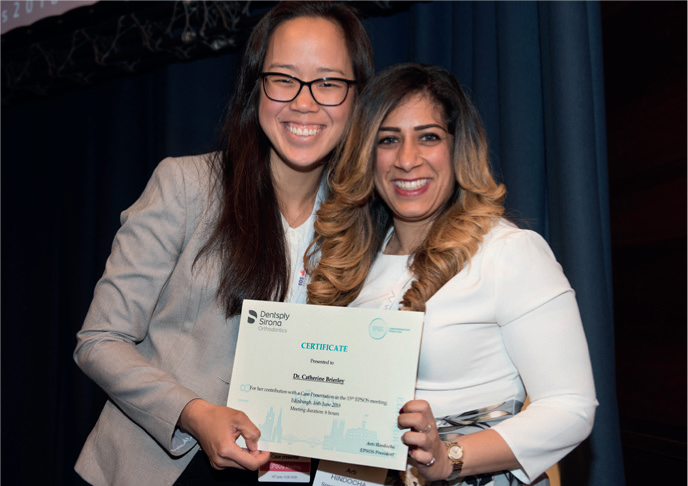
The examination also coincided with the EPSOS (European Postgraduate Student Orthodontic Society) meeting, which was free of charge. This provided me with the opportunity to complete my examination and do an oral case presentation at the EPSOS conference. Overall, I had an extremely positive experience at both the provisional EBO examination and the EPSOS meeting (Figure 1).
Figure 1. Catherine receiving her certificate from Arti Hindocha in recognition of her case presentation at the European Postgraduate Student Orthodontic Society Conference.
Full EBO membership – Trevor
It was back in 2014 in the company of Jon Sandler, Julian O'Neill and Ewa Czochrowska (all EBO diplomates) that I got the idea of working up cases for the EBO. As an independent specialist of at least five years, the only route available was to work up eight cases with retention records, and also sit the oral examination described above. Fortunately, working in both the primary and secondary care settings resulted in my having a wide range to my clinical practice, which helped enormously when having to select cases.
Reasons for applying
As a ‘goal orientated’ person, I was looking for a new professional challenge and, having been established for a number of years, both in practice and hospital, I thought that this would be a real clinical challenge. I set out to search prospectively for appropriate cases to work up and also intentionally record, in a database, patients whose records I could subsequently use as teaching material, which involved taking significantly more clinical photographs than previously.
Preparation for examination
The guidance is very clear as to the description of the eight cases you are required to submit for full membership, along with an oral viva of two unseen cases.5 I did not rule out any patient that fitted a case category, making sure that I had a few in each category to choose from, just in case of untoward events. Taking additional photographic records and closely guarding study models took me back to ‘MOrth days’ more than a decade earlier.
Undoubtedly, there were times over the last few years when I questioned the wisdom in pursuing membership of the EBO, but being a ‘completer finisher’ meant quitting was not an option …and finally I had my eight cases (Table 2). I was particularly pleased that the selected cases reflected my three main areas of practice. Case 2 had been treated on a ‘fee paying’ basis, Case 5 had been undertaken in the hospital service and the remaining six cases had all been treated under the National Health Service in a primary care facility.
1. EARLY TREATMENT MALOCCLUSION
Class III malocclusion in mixed dentition with agenesis: UR2 & UL2Stage 1 – URA to correct anterior crossbiteStage 2 – Fixed appliances non-extraction
2. ADULT MALOCCLUSION
Adult patient with Class I malocclusionCrowding and upper centreline shift to leftExtraction of UR4, LL5 & LR5Fixed appliances
3. CLASS I MALOCCLUSION
Class I malocclusionCrowding with lower centreline shift. UR5 & UL4 in scissors biteExtraction of UR6, UL6, LL6 & LR6 and fixed appliances
4. CLASS II DIVISION 2 MALOCCLUSION
Class II division 2 malocclusionRetroclined upper incisors, increased overbite and Class II canine relationship on rightExtraction of UR4 & UL5 and fixed appliances
5. CLASS II DIVISION 1 MALOCCLUSION
Class II division 1 malocclusionSN to Go-Gn angle of 49°Extraction of LL4, LR4 & LR8 after previous extraction of UR4 & UL4Fixed appliances and Orthognathic Surgery
6. CLASS II DIVISION 1 MALOCCLUSION
Class II division 1 malocclusionOverjet 14 mm and LL5, LL6 & LL7 in scissors biteTwin block functional applianceExtraction of UR5, UL5, LL5 & LR5Fixed appliances
7. A SEVERE SKELETAL DISCREPANCY
Class II division 1 malocclusionOverjet 19 mm A-N-Pg angle of 5°Twin block functional applianceFixed appliances non-extraction
8. A SIGNIFICANT TRANSVERSE DISCREPANCY
Class I malocclusionOverjet 2 mmBroad lower arch: UR6, UR5, UR4 & UR3 in crossbiteQuadhelix and fixed appliances non-extraction
The preparation guide suggests that it takes about 12-15 hours to prepare for each case. In my experience, this was probably an underestimate, so starting early was essential. It is also a good idea to have a look over a previous diplomates cases.
The examination
Although there were no time constraints, I was keen to sit the Edinburgh 2018 diet, partly because I had previous success in Edinburgh in 2002 sitting MOrth, but mostly because it was easier to get on a train at Leeds with 25 sets of plaster models to take to Edinburgh, than travelling to some exotic, but far-flung European destination.
Although it had been 14 years since I had an examination, I have to say I was not unduly worried about being examined on unseen cases. I was, however, extremely nervous about my ‘finest cases’ being scrutinized by experts with a fine toothcomb – these really were the best I could do. An example of the standard expected is illustrated below. This case fitted the fifth category – a Class II division 1 malocclusion with a high Frankfort mandibular plane angle (minimum 30°) and/or SN to Go-Gn angle of 37°.
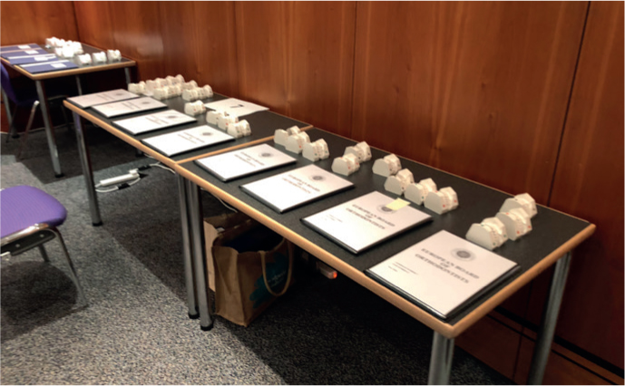
In the event, when I was called in to receive my feedback only 24 hours after the exam, I was delighted to be told I had passed. At this stage the examiners had critiqued every single detail of all the cases presented (Figure 2). I was impressed with their attention to detail and the time they had taken examining my cases. I was also delighted that they chose one of my cases (Case 7) to put on display for the benefit of future EBO candidates.
Figure 2. The eight cases laid out for examination for full membership.
Example EBO case. Case 5 – Class II division 1 with high FM angle (TH)
A 22-year-old male patient presented complaining of his top teeth ‘sticking out’ and a ‘short lower jaw line’. The patient had been referred following a previous course of orthodontic treatment with functional appliances and then the loss of both upper first premolars and fixed appliances. He had previously sustained uncomplicated crown fractures on the mesial aspects of the upper central incisors aged 7 years, but these teeth were responsive to thermal stimulus. Whilst the patient was aware that there was some staining around the composite restorations placed on the palatal aspects of these teeth, he was not requesting replacement.
Examination
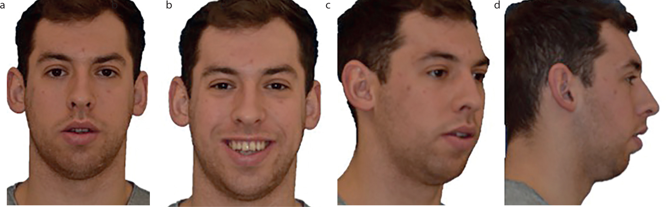
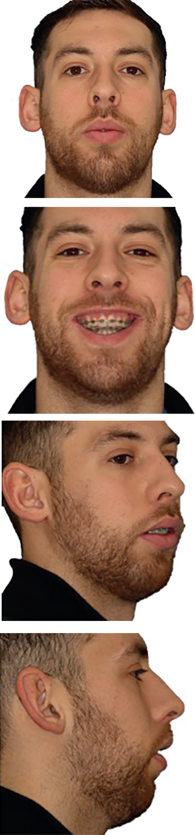
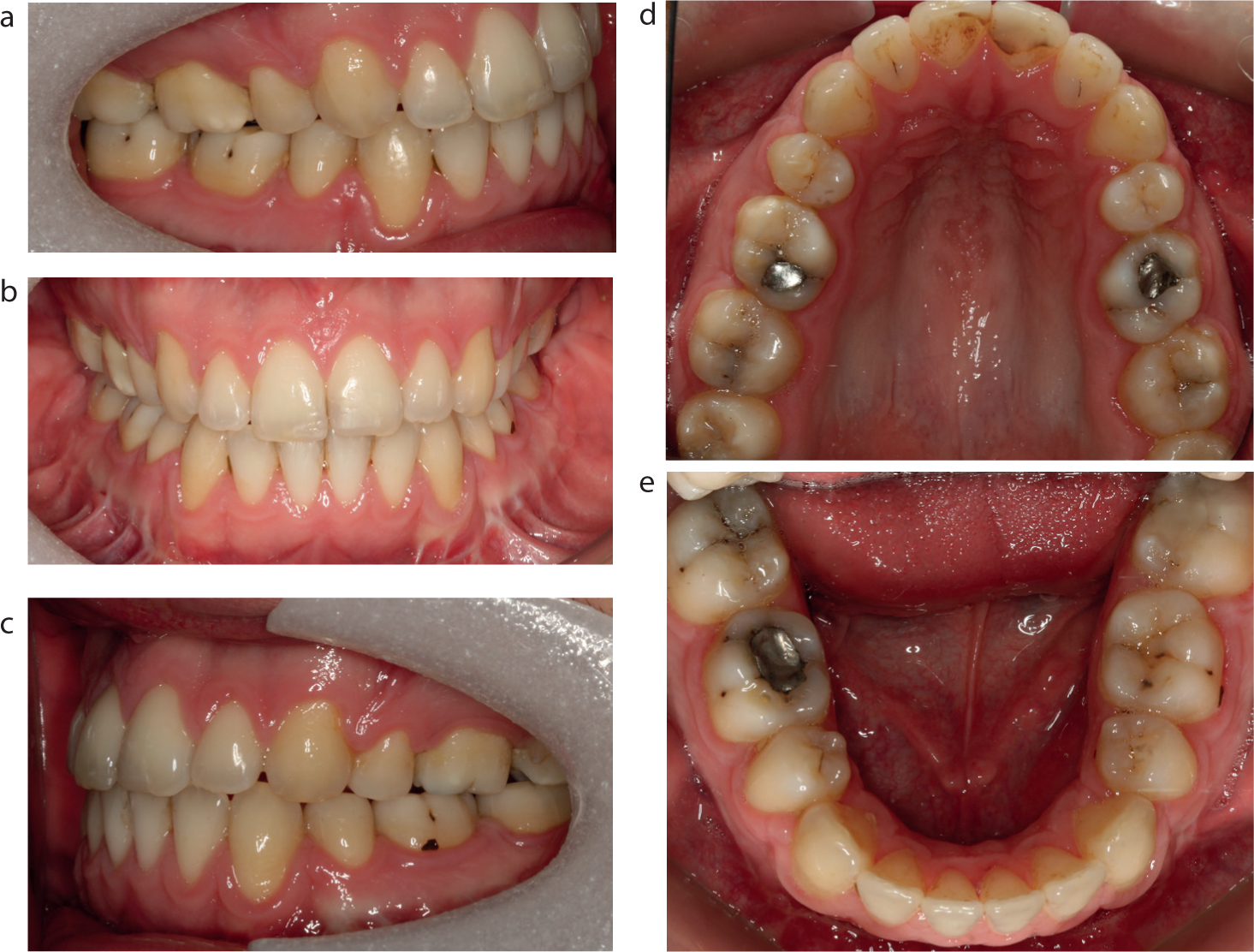
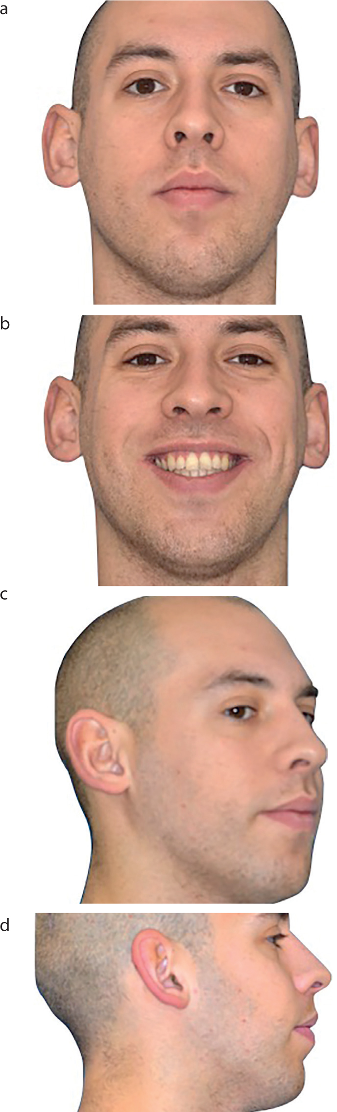
Examination revealed a Class II division 1 incisor relationship on a severe skeletal II base with mandibular retrognathia. The Frankfort–mandibular planes angle and lower face height were both increased. The lips were incompetent at rest (Figure 3). There were no signs or symptoms of TMD. Intra-orally the oral hygiene was good.
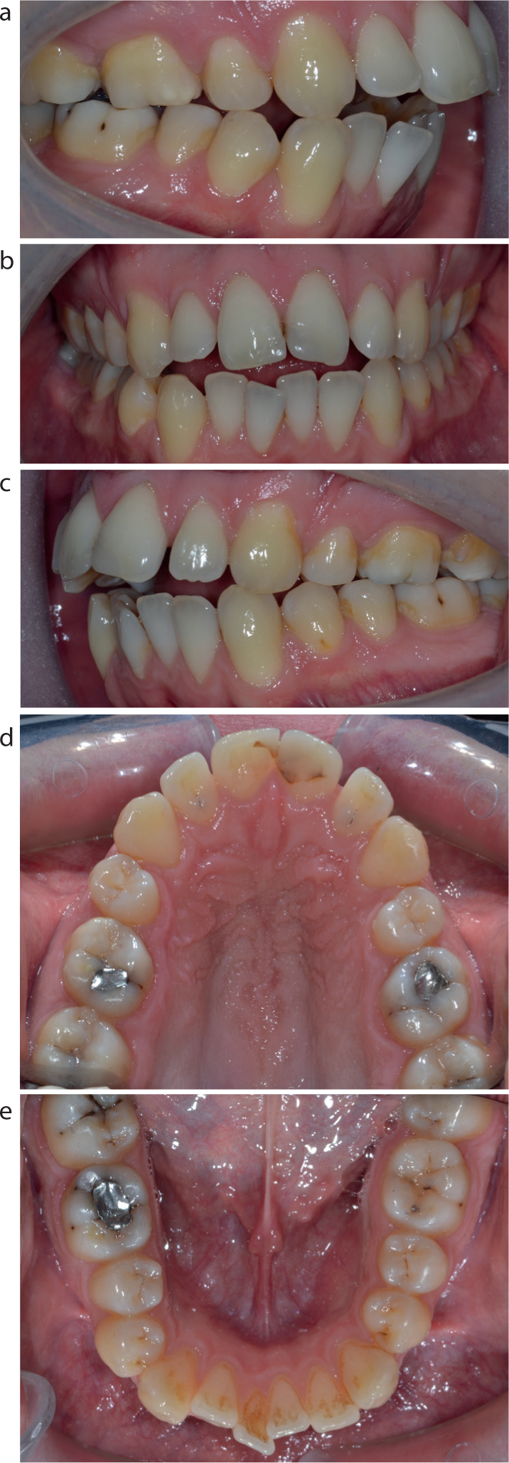
In the maxillary arch, the labial segment was spaced and the incisors were proclined. In the mandibular arch, the labial segment was mildly crowded and the incisors were proclined.
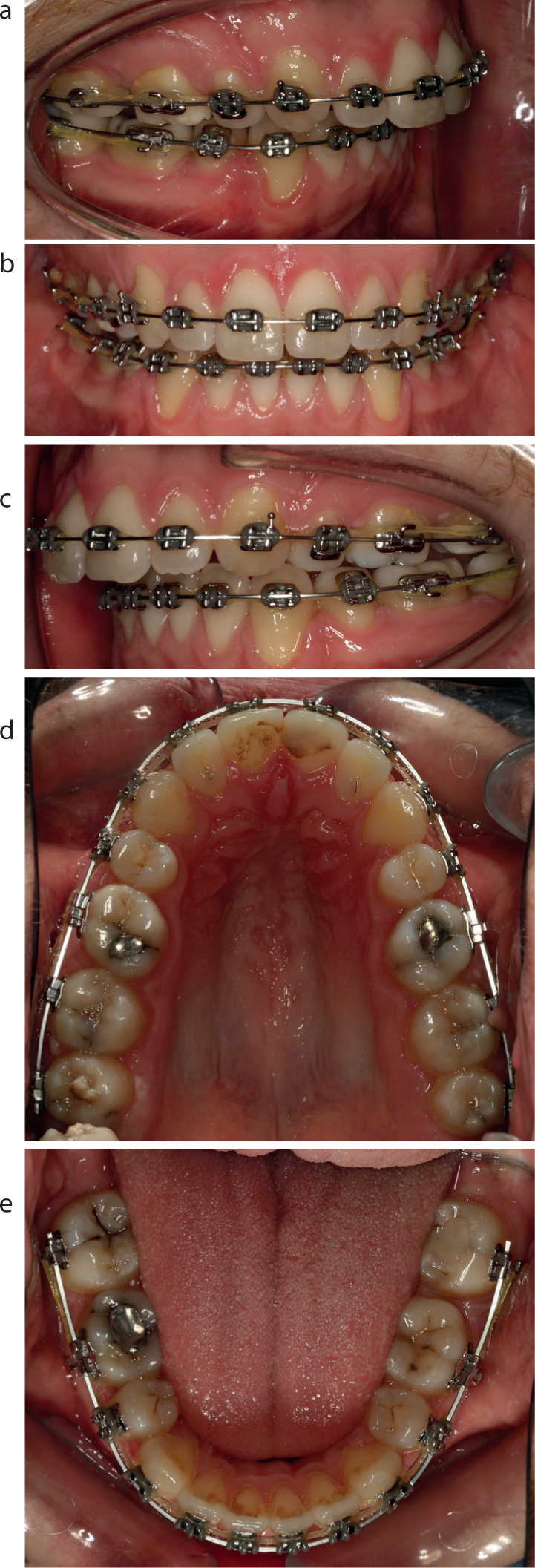
In occlusion, there was an overjet of 8mm and there was an anterior open bite extending to the upper second premolars. The lower centre line was 2 mm to the right of the facial midline. The molar relationship was a full unit Class II bilaterally whilst the canine relationship was a ¾ unit Class II on the right and a ½ unit Class II on the left (Figure 4). On both right and left excursion there was group function and the retruded contact position and intercuspal position were coincident.
The Dental Health Component of the IOTN was 4a and the Aesthetic Component was 8.
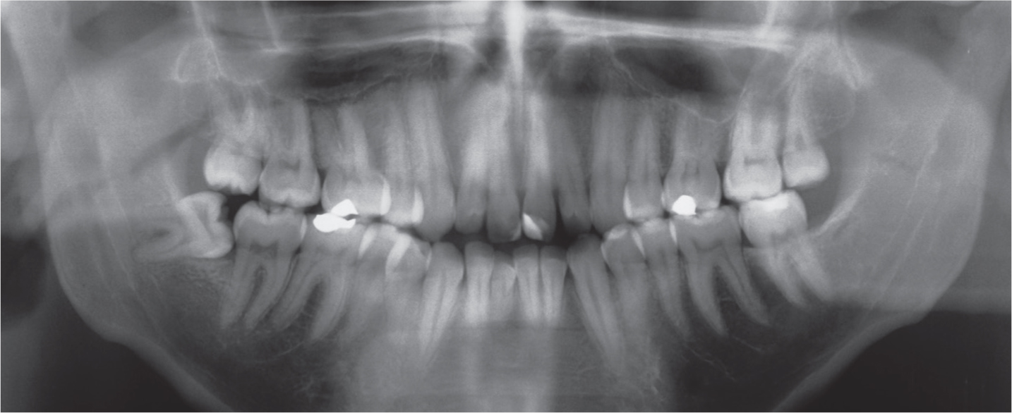
The OPG radiograph showed all the permanent teeth to be present with the exception of both upper first premolars and the lower left third molar. No carious lesions were detected, the root morphology was normal and the alveolar bone levels were good (Figure 5).
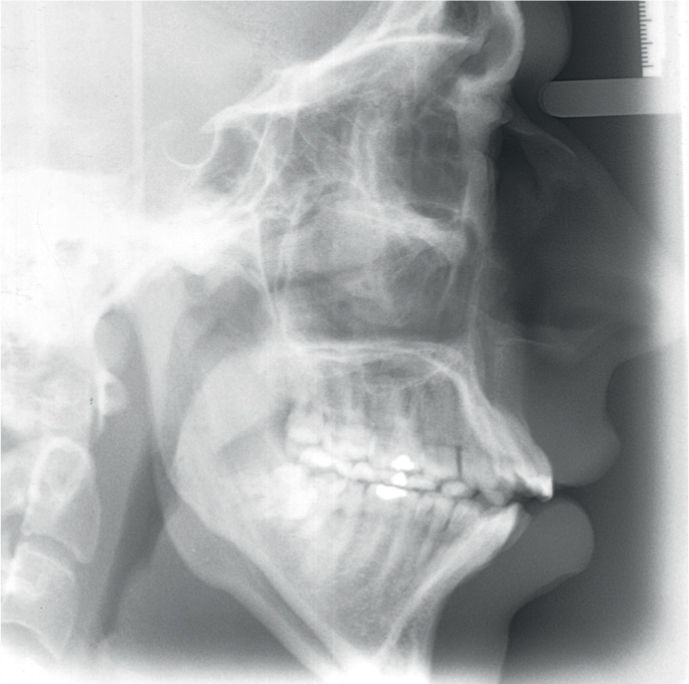
Cephalometric analysis confirmed the clinical findings of a severe Class II skeletal pattern with an ANPg of 10°. The maxillary–mandibular planes angle at 38° and the SN to Go-Gn angle at 49° were both increased. The upper incisors were proclined at 120° and the lower incisors were proclined at 107°. The lower incisors were 11 mm ahead of the A–Po line. Figure 6 shows the pre-treatment lateral cephalogram and the cephalometric analysis is presented in Table 3.
Sagittal and vertical correction of the malocclusion to improve the facial profile;
Align the teeth and correct the incisor relationship with reduction of overjet and achievement of Class I molar and canine relationships;
Achieve a good functional and static occlusion.
The treatment plan involved:
Extraction of lower first premolars to allow for decompensation of the lower incisors and extraction of the lower right third molar to aid surgical cuts;
Provision of upper and lower edgewise pre-adjusted fixed appliances to level and align the teeth, close the lower arch spaces and upright the lower incisors;
Bimaxillary orthognathic surgery to impact the maxilla and advance the mandible;
Genioplasty to advance the chin point;
Detailing and finishing of the occlusion;
Debond;
Retention with vacuum-formed removable retainers in both arches.
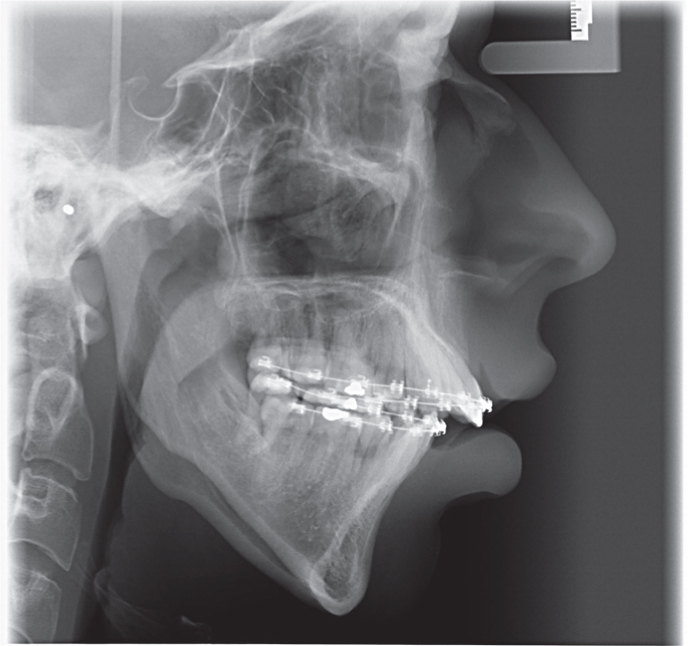
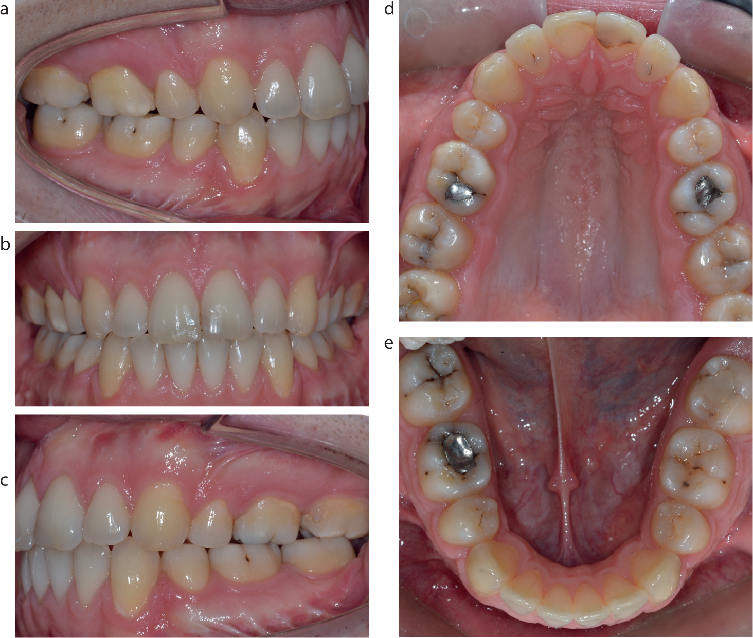
Following the extraction of the lower first premolars and the lower right third molar, full arch fixed appliances were then placed with the insertion of 0.014” Sentalloy archwires. After 14 months of alignment and space closure on 0.019 x 0.025” stainless archwires, impressions were taken to check on co-ordination of the arches in preparation for orthognathic surgery. Full records were then taken (Figures 7 and 8), including a lateral cephalogram, which showed correction of the upper incisor inclination, decompensation of the lower incisors, thereby increasing the overjet, and an improvement in the overbite ahead of surgery (Figure 9). In discussion with the patient and the maxillofacial surgeon, a surgical plan was agreed for a non-differential maxillary impaction of 3 mm, a maxillary advancement of 2 mm, a bilateral sagittal split osteotomy mandibular advancement to fit and an advancement sliding genioplasty.
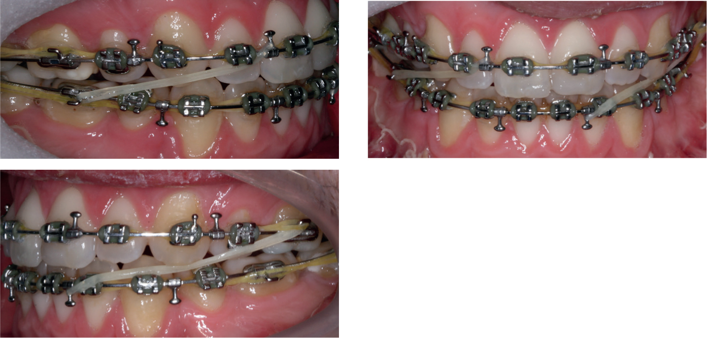
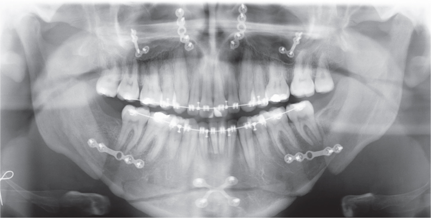
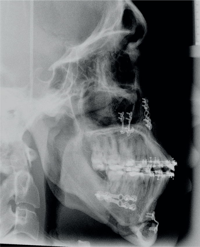
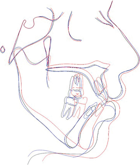
Post-surgery the patient was reviewed at two weeks (Figures 10 and 11) and inter-arch elastics (3.5 ounce, ¼”) were introduced (Class II on the right and Class III on the left) to settle the occlusion. At one-month post-operation, a dental panoramic and lateral cephalometric radiograph were taken for analysis of the changes achieved (Figures 12 and 13). The dental panoramic radiograph showed good root parallelism, bone levels and root lengths and no pathology. The final lateral cephalogram showed the desired maxillary impaction and mandibular advancement had been achieved, although the advancement sliding genioplasty could have involved a greater A-P movement. The maxilla and mandible positions were now within the normal range and, whilst the skeletal base relationship has remained Class II and the vertical dimensions increased, these were both much improved from the start of treatment. These changes are illustrated in the cephalometric superimpositions shown in Figure 14. Nineteen months after the start of treatment, the appliances were debonded and upper and lower vacuum-formed retainers were fitted (Figures 15 and 16).
Figure 10.
(a–d) Post-surgical extra-oral views.Figure 11.
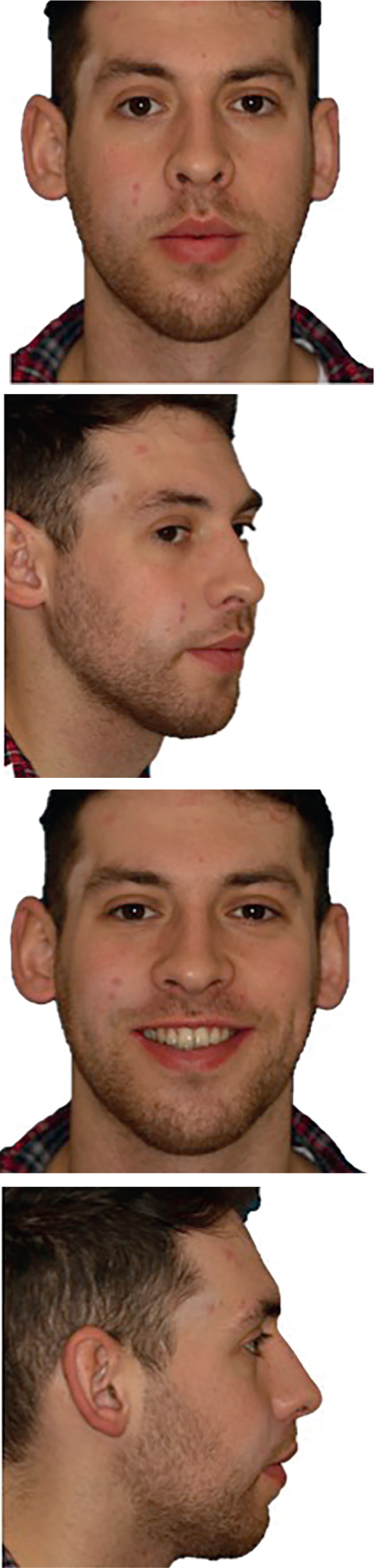
(a–c) Post-surgical intra-oral views showing settling box elastics.Figure 12. Dental panoramic radiograph near end of treatment.Figure 13. Lateral cephalogram near end of treatment.Figure 14. Superimposition on SN line of pre-treatment (black), pre-surgical (blue) and end of treatment (red) lateral cephalograms.Figure 15.
(a–d) End of treatment extra-oral views.Figure 16.
(a–e) End of treatment intra-oral views.
Overall treatment saw the profile improve significantly as a result of bimaxillary surgery. The upper lip projection has improved, the lips are competent, the chin point projection has improved and the chin-throat angle has improved, although there could have been more upward and forward slide of the genioplasty. In excursion, canine guidance was evident on the right and group function on the left, with no non-working side interferences. The retruded contact position and the intercuspal position were coincident. The alignment and space closure achieved resulted in a good final interdigitation on both sides. The pre-treatment PAR score of 43 had reduced to 2, representing a 95.3% reduction.
At a 2-year debond review it was evident that the profile and the position of the teeth following treatment had been maintained and these were still providing good lip support. The dental alignment and occlusion had remained stable, with good compliance with the retention regimen, and the patient was delighted with the result (Figures 17 and 18)
One of the objectives of the EBO is to enhance the standard of orthodontic treatment throughout Europe by providing a standard against which the orthodontists, who so desire, can be judged independently of national examinations and barriers. In a time where our own Royal College specialty examinations are changing, and the emphasis on submitting treated cases is perhaps diminishing, maybe more people in the UK will choose the EBO as a professional goal with clinical relevance, for their own improvement and possibly for the benefit of the patients for whom they care.