Article

Our assessment of the aetiology of the malocclusion was that the dento-alveolar segment was positioned too far back on the basal bone of the mandible. We wanted, therefore, to correct the 12-mm overjet and the Class 2 dental occlusion, but to have minimal effect on the chin position because we found it to be acceptable pre-treatment. We decided to leave the lower border of the mandible alone and to do a total subapical osteotomy.
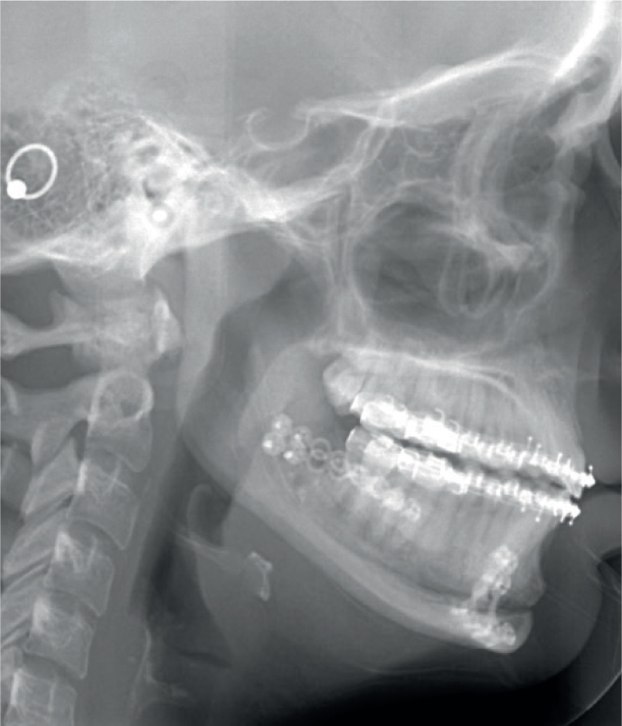
This procedure was first described in the literature by MacIntosh in 1974,1 and further successful cases have been documented.2 This procedure was thought to be most appropriate in cases where a change in the lower labio-mental sulcus was needed, and it has the advantage that the entire dento-alveolar segment can be advanced en bloc. However, the chin point remains exactly where it started, thus avoiding the need for a genioplasty, the results of which are unpredictable (Figure 1).
Potential disadvantages of this approach include the possibility of sensory nerve damage, loss of tooth vitality and the time required to complete this meticulous surgery.
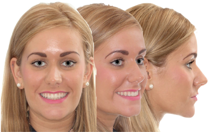
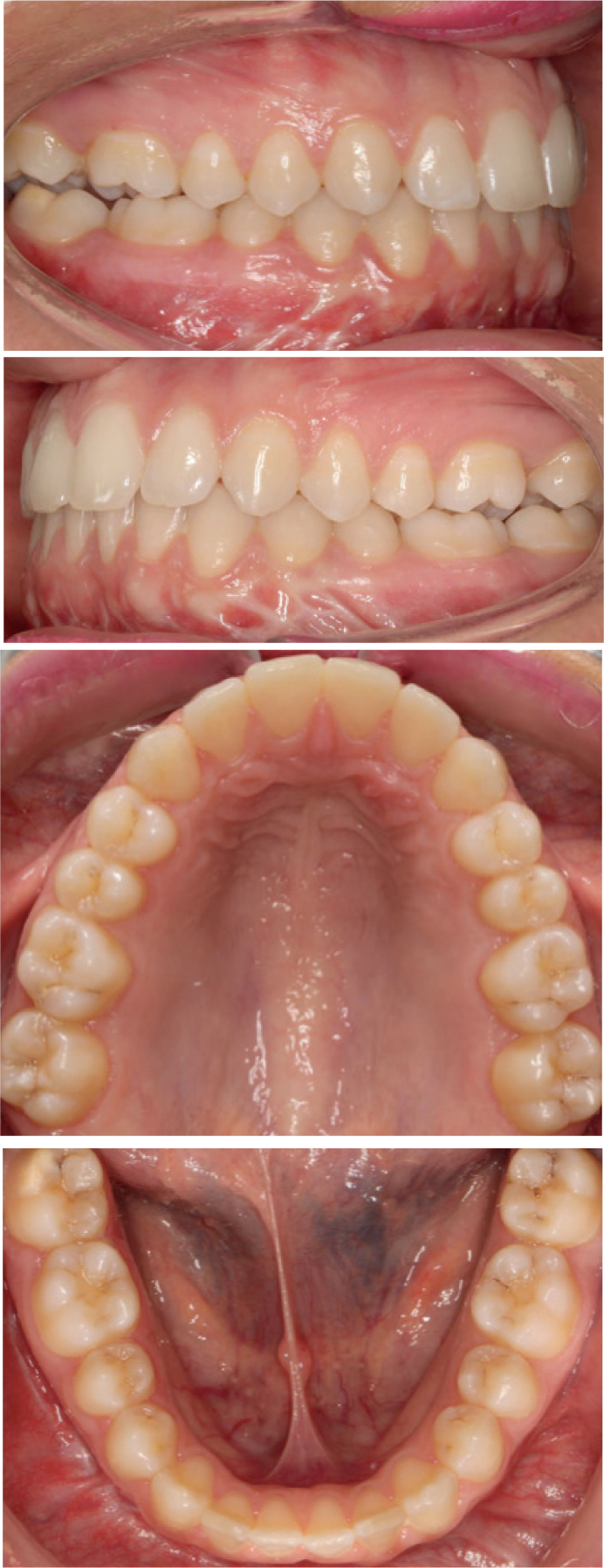
In this case the patient was delighted with the facial results achieved (Figure 2) and was thrilled with her improved occlusion (Figure 3). At a 12-month post-debond review, the patient reported no significant nerve deficit.